


Editor’s note: We are delighted to present this excerpt from Chapter 3 (“A Voyage Through the Cell”), in the new book Epigenetics and the Architect: Evidence of Design at the Frontier of Biology, by Thomas E. Woodward and James P. Gills, MD (Discovery Institute Press).
Imagine you have been invited to a futuristic discovery center, a lavishly funded facility that has pioneered the ability to shrink people and objects many orders of magnitude. A wild fiction, to be sure. Even if such miniaturization were possible, any number of fine-tuned parameters of the laws and constants of physics and chemistry that make organic life possible would be thrown into havoc by the miniaturization. We would arrive on the other side decidedly dead. But that never stopped Ant-Man or the Magic School Bus or the Szalinski family in Honey, I Shrunk the Kids or the crew of Innerspace or, for those readers with even longer memories, The Incredible Shrinking Man or the crew of Fantastic Voyage. So, let’s climb into the incredible shrinking submarine with our tour guide and begin the journey.
The View from Inside
The strange vessel is equipped with panoramic windows in every direction. As you buckle in, everyone is encouraged to close their eyes. A launch countdown sounds over the intercom. You feel a bit of vertigo as if a recklessly fast elevator were speeding downward from the top level of a skyscraper. There’s a shrill metallic ringing and then both the sound and all sense of motion stop. A moment later you’re jerked back into motion, but now the motion feels like that of a ship in roiling waters. Your piloting tour guide, seated with you and the other members of the tour in the spacious cockpit, raises a hand to signal that all is well. “They’re moving us into position,” he explains, “just a few billionths of an inch from the water-engulfed surface of a living cell.”
The jerky motions of the ship give way to a steadier, more stately motion. “Okay,” your tour guide says, “go ahead and open your eyes.”
When you do, there before you, on the other side of the submarine’s main viewing window, is an enormous cell — or at least it appears enormous, since you and the submarine have been shrunk down to a tiny fraction of the cell’s size.
The vessel approaches the outer surface of the cell and slips into one of the many gates in the cell wall. Now all around you are the bobbing heads and trailing fronds that form the cell wall’s double-layer lipid membrane. After squeezing through this dark curtain-like structure, your vessel passes into the cell interior, marked by a lattice-like maze of beams and girders. It looks as if some gargantuan and enormously intricate skyscraper were under construction beneath the sea — with all the interior pillars, trusses, and girders laid bare in a vast underwater cavern.
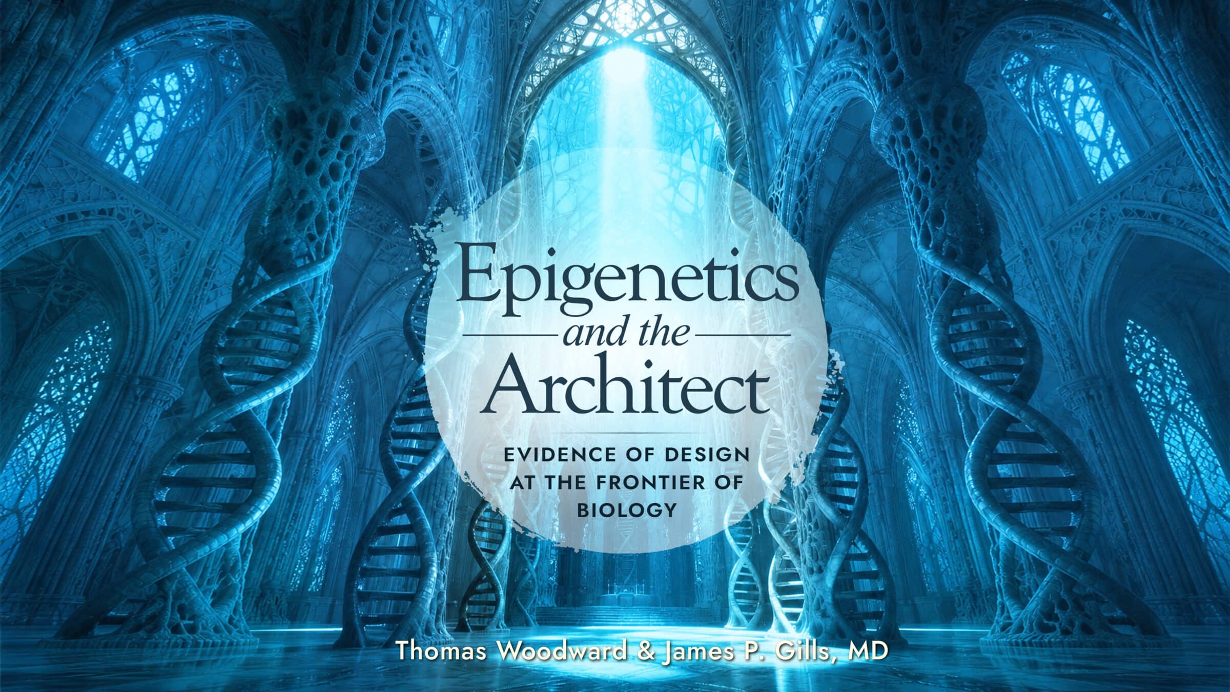 Image source: Discovery Institute Press.
Where the Epigenetic Information Resides
Your guide gestures with a sweep of the hand. “What you see all around you is the internal structural skeleton of the cell. Most of you are familiar with DNA, with genetic information. We’ll encounter plenty of that. But some of the cell’s most important epigenetic information is embedded here, in the precise way that these structural girders are positioned and attached to the cell membrane. This informational pattern is especially important in a newly fertilized egg — the zygote. Even when it is still just a single undivided cell, the zygote has far more information in its 3D structure than we imagined, far more than just what is rooted in its DNA.”
After descending through a maze of tubular braces and beams, you spy ahead the nucleus, the spherical storehouse of the cell’s DNA. At the tiny scale you and the sub have been shrunk to, it looms as a huge, shimmering globe, an underwater city whose surface teems with the traffic of complex molecules shuttling to and from its surface. As the sub draws closer, you see that the molecular blobs pass in and out of the nucleus via hundreds of large circular pores in its wall. Each portal has tiny hairs protruding around the entrance. The submarine approaches one of these portals and slips through.
Arrayed before you is a vast and crowded underwater arena. Hundreds of aisles penetrate the maze of stacked loops and clumps of DNA in every direction. Your tour guide pilots the sub into one of these narrow aisles and aims the sub’s directional searchlight on a wound-up mass of DNA.
“You know those spools in sewing boxes, with the thread around them?” he says. “Here the DNA is the thread and the histone complex is the spool — only, as you can see, it’s a lot less tidy than a sewing-box spool. Don’t sell it short, though. It’s an engineering wonder, enabling DNA to be compacted to an extraordinary degree. Each histone protein — reminiscent of a tiny clump of pasta — is a string of one hundred or so amino acids precisely folded into an irregular Z-shape. Once folded, eight of these histone proteins combine like a three-dimensional jigsaw puzzle to form the ideally shaped spool, called a histone core. In human cells millions of histone cores are produced as the cells divide and stand ready for the new DNA to be wound around them.”
Something Odd Sticking Out
Your pilot tour guide points the searchlight at something odd sticking out of a spool on one side. It looks vaguely like a thin branch, or a tail. As the sub draws closer, the pilot illuminates three other tails like the first one. “Each histone core is equipped with eight tails. You can spot four of them on the side facing us.”
He narrows the spotlight beam and trains it on a strange Y-shaped tag attached to one of the tails that seems to be bulging out and pulling away from a histone bundle. “Note what is happening here,” he says. “The little molecular Y-tag is causing the whole tail to be steadily pulled away from the layers of DNA on the spool. This is a key marker in the epigenome system. The tag is an acetyl molecule, attached to a precise spot on the projecting tail of the histone spool with the help of a special protein machine. This particular tag functions as an unlocking device. When the acetyl tags are placed on the tails, those tails swing away from the spool, and this makes it much easier for the file of DNA that is wound onto that histone to be pulled off and read. On the other hand, if the acetyl tags are removed by a different protein machine, the slender histone tails snug up closer to the DNA wrapped around the spool, allowing the tail to act like a clamp, securing the DNA tightly onto the spools and making it harder to be read. It’s like a locking and unlocking system on a filing cabinet. These acetyl-tagged histone spools are all over this DNA warehouse. If we tried to count them all, we’d be here a long time. The information required to manage the precise placement of acetyl tags, something like a dynamic 3D map, is stored beyond DNA. It’s epigenetic.”
Complicated Enough for You?
Outside the submarine window, a protein machine suddenly swoops down and removes an acetyl tag from the spool. “That’s a histone deacetylase enzyme, or HDAC, engineered to grab and remove tags,” the pilot says. “It’s complementary protein machine, a histone acetylase enzyme — HAC for short — attaches the acetyl tags. If all that is not complicated enough, there are four other chemical tags also found on those tails.”
You sense a hum and a low vibration. The intensity builds steadily, and then there’s a voice over the intercom. It’s mission control. “Incoming. Take evasive maneuvers.”
“Roger that,” says your guide, and deftly maneuvers the submarine away from the DNA spool.
The ominous vibration persists, but then a broad smile breaks across your guide’s face as he looks toward the far end of the narrow DNA corridor. “We’re in luck,” he says. “We happen to be in a section of Chromosome 17 where a key stretch of DNA is being opened and read. The vibration you’re feeling is the rumble of the big protein machines lumbering along the stacks of DNA just ahead. They’re unlocking and prying open very specific sections of the DNA spool clusters. We call these clusters nucleosomes. The goal of the machines is to gain access to the target cluster of genes.”
The pilot seems to have forgotten all about the command to take evasive maneuvers. Instead, he aims the sub directly into the action, a frenzy of strange underwater robots scurrying around the tiny, interconnected islands of DNA. One slice of the action stands out. As the genetic corridor opens wider, several DNA-manager bots swarm down on the DNA, grabbing, twisting, and rapidly unwinding sections of it from their spooled-up position on the histones. Soon they have the DNA molecule unfurled.
Like Fuzzy Yarn
At first, the double helix up close looks like fuzzy yarn, but as the sub draws closer, the DNA’s elegant spiral-ladder geometry comes into focus. Its unique form looks like what is depicted in textbooks, except the rungs are knobby and thick, with almost no space between them.
The tour guide pilots the sub down along the spine of the DNA, and as they pass by, you get a close-up of the smooth surface of the DNA letter pairs, the rungs in the twisting ladder. Your guide aims the spotlight at one of the rungs. “Notice here and there, the smoothness of the rung pattern is marred by something sprouting from the rung — a tiny antenna-like appendage resembling a baby sprout of broccoli. No, it’s not an unfortunate growth. The molecule is a chemical marker called a methyl tag. In its free state, as methane, it has five atoms — one carbon and four hydrogen. But the methyl group has one less hydrogen atom — three instead of four. So think of the methyl tag as a stripped-down methane molecule — a minor variation on the gas molecule burned by the trillions in any stove fired by natural gas.
A Core Feature of the Epigenome
“Researchers discovered this tag in 1948, and they learned that it gets attached only to the C letters in DNA’s four-character alphabet, and only to some of them. Methyl tagging is one of the core features of the epigenome. Tens of millions of DNA rungs are tagged this way, and it was discovered that this tagging pattern is indispensable to cellular function. The specific letters getting tagged are chosen for functional reasons. Some molecular biologists describe the tagged Cs as the fifth letter of DNA. On this view, there is A, T, C, G, and methylated-C. That last letter is crucial. Typically, it acts as an off switch for the stretch of DNA where it’s attached. If all those stretches were on all the time, we could expect a nasty multi-molecule pile-up. Some scientists estimate that as many as 200 million methyl tags have been placed on precise locations on the DNA in a single nucleus.”
The tour guide checks the sub’s instrument panel and then resumes his commentary. “When a cell divides, the entire genome is opened up to be duplicated. All the billions of rungs of DNA are split open and replicated. So, do all these methyl tags get copied into the bargain?”
No one volunteers an answer beyond a reticent nod of the head from a fellow passenger.
A Special Protein Machine
“Exactly!” your guide replies, seizing on the weak gesture of assent. “The methyl tags are also copied over to the same corresponding rung in the new cell’s DNA. The ‘how’ is interesting. The job falls to a special protein machine — a methyltransferase. But there’s a catch. The pattern of methyl tagging varies from cell to cell, and while this methylation map is generally passed on unchanged from mother cell to daughter cell, there are untold thousands of exceptions, especially when an embryo is developing. The differences are essential, and the source of the differences isn’t limited to DNA.”
He checks the instrument panel again and then straightens in his chair. “Oxygen supplies getting a bit low. Time to make tracks.”
If the lazy tune he begins whistling is any indication, he’s expecting an uneventful return journey. But after your vessel passes through the lipid bilayer membrane, it is only past the cell wall for a moment before a second cell, an angry-looking bacterium, bears down on your ship.